

Histalim is a food histology laboratory that has been offering histological analysis on meat products, truffles, foie gras and fish since 2005. We are the only laboratory in France accredited by COFRAC to ISO 17025 norm since 2006.
For non-food related studies, please go to Cerba research
Histology of composition – COFRAC Accredited
We provide solutions to determine whether your meat and seafood products comply with the applicable regulations and to assess their quality. We can intervene with both raw materials and finished products.
In a single analysis using a microscope, our analyst lists the ingredients and additives, delivering a conformity statement for the sample.
We can:
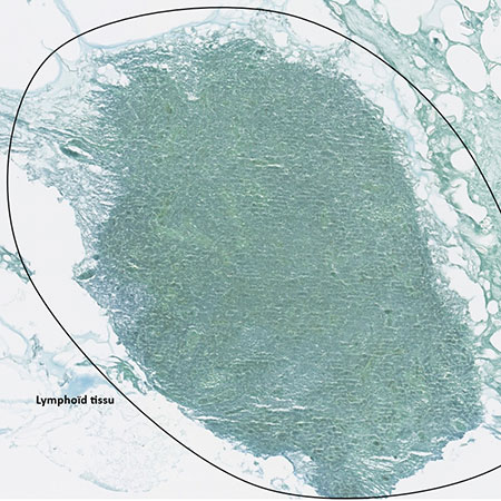
- Detect the presence of certain tissues (glands, lymphoid tissue…)
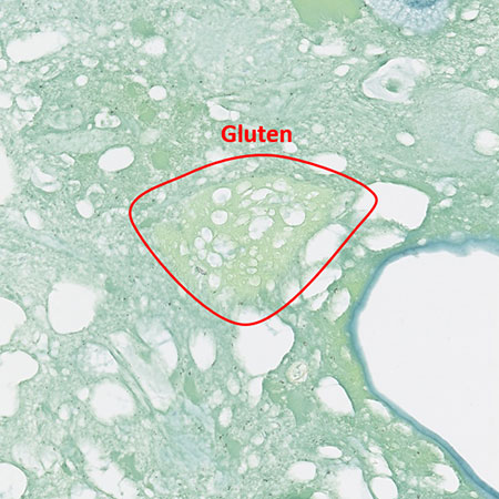
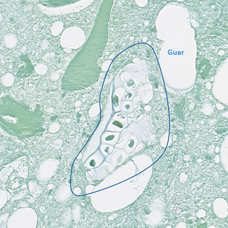
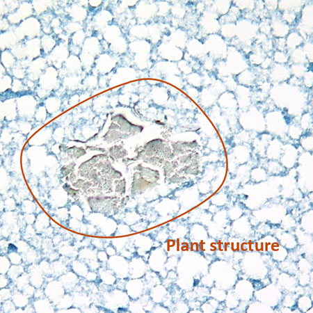
- Detect the presence of additives (gelling agents, plant proteins…)
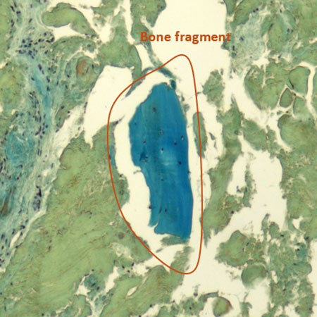
- Detect the presence of Mechanically Separated Meat (MSM)…
We are able to differentiate more than 70 elements.
Chemical and DNA analyses do not allow the determination of various tissues contained in a preparation or raw material, unlike histological analysis.
Pâté sample
Chicken nuggets sample
Sausage sample
Minced beef sample
Analysis on Truffles
We propose two types of analysis to check the quality of your canned or fresh truffles, preparations, truffle cracks, peels or juice:
- Identification of truffle species (A): We can identify 7 different truffle species, including Tuber melanosporum, Tuber aestivum, and Tuber brumale, even in the case of truffle-based products (butter, cheese…). This analysis allows us to determine if your product is composed of the declared truffle species and if it is subject to Decision No. 95 of CTCPA.
- Search for exogenous materials (B): We can indicate the presence of gelling agents (if insoluble in water and/or fat) as well as parasites often found in truffles.
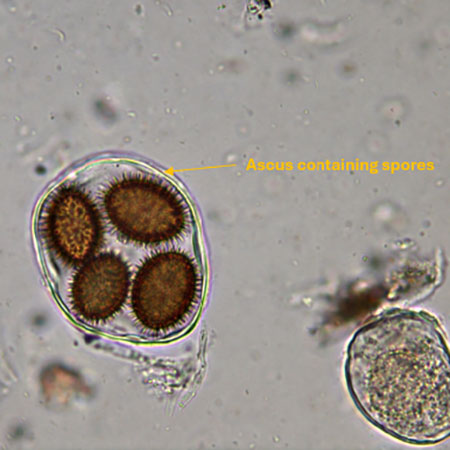
(A): Tuber brumale truffled butter
Identification of truffle species
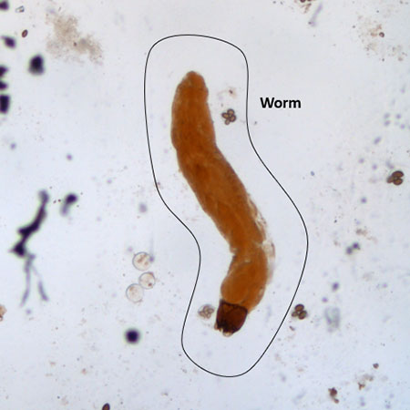
(B): Tuber melanosporum truffle juice
search for exogenous material
Foie gras
If you want to evaluate the quality and compliance of your whole foie gras samples or preparations, our histological analyses can meet your needs.
Histology of composition analysis (COFRAC accredited) (A):
This analysis follows the principle of histology of composition analysis. It can indicate the conformity of your product with the criteria of French decree No. 93-999 of August 9, 1993, related to preparations based on foie gras and/or the list of ingredients declared on the labelling.
Fattening level (B):
Through this analysis, we can observe various pathological changes (fibrosis…) as well as the liver fattening level. The conclusion of this analysis can be made according to the French decree of April 8th, 1994*, related to official methods of analysis of preparations based on foie gras.
*Conformity with other specifications can be studied upon request.
(A): Foie Gras block sample (Histology of composition)
(B): Whole Foie Gras sample (Fattening level)
For any further information or a detailed presentation of our analysis catalog, please contact us
Histalim processes your data in order to contact us.
To find out more about how we manage your personal data and to exercise your rights, please refer to our Privacy policy.